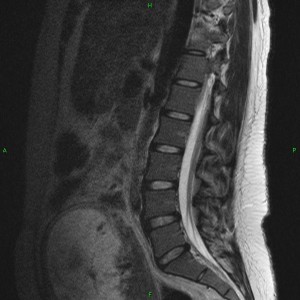
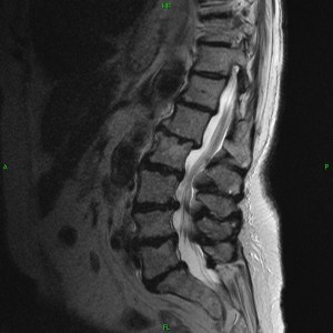
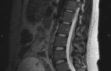
In young healthy individuals, there is a straight alignment from one lumbar vertebra to another, the edges of each vertebra are uniform and regular in appearance, the disc spaces are well maintained (not flattened), the disc walls are straight (not bulging), and the color of the discs on MRI imaging is white, signifying a healthy level of water hydration within the discs. With aging, and as physical loads are applied to the spine, the alignment of the spine shifts, the discs lose their hydration (now appearing dark on MRI), and the disc walls begin to bulge or herniate – typically along the posterior disc wall margins (from repeated forward flexion at the waist). The pictures shown below highlight some of the typical MRI appearances associated with this progression. The vertebrae are the square shaped gray structures stacked in a column, the discs are the small band shaped structures between each vertebrae, the while vertical tube to the right of the spine is the spinal column where nerves travel, and the spinal cord is the grayish vertical stranded structures traveling within the canal. As you can see, spine deterioration can cause crowding of the spinal cord (nerves) within the bony spinal column. These are views of the lower back seen from the side or lateral viewpoint.
Young Spine:
Middle Aged Spine:
Old Spine:


